


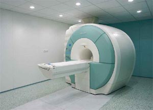
Its short name is MR or MRI or MRI and it is pronounced Emar. It stands for Magnetic Resonance Imaging. Magnetic Resonance Imaging (MR) is a painless diagnostic technique that does not require medication to cause allergies and does not use harmful tools such as x-rays.
SPECIAL MARKETING EXAMINATIONS
Emar MR Arthrography
It is a diagnostic method that provides detailed visualization of intra-articular structures. In this way, structures that are difficult to see in normal MRI examinations are visualized. Contrast substance mixture is injected into the joint with a thin needle to cause swelling in the joint capsule. After a short waiting period, MR examination is performed. Injection is performed in some joints under the guidance of US or scopy.
Emar MR Urography
MR Urography is a non-invasive imaging method used to view various urinary system pathologies in the clinic. MR urography is preferred over conventional methods in pregnant children and patients with contrast material allergy or renal failure.
Emar MR Cholangiography
Cholangiopancreatography (MRCP) is a non-invasive examination technique with proven reliability in the evaluation of the pancreatic and biliary system. Thus, stones belonging to the biliary tract, tm. It helps in the diagnosis of diseases such as
Breast MRI
Unlike mammography and ultrasonography, in addition to the morphological features of breast lesions such as shape, contour and size, it shows the tissue perfusion characteristics of the breast parenchyma and mass lesions on this background. The ideal period for breast MRI is between the 5th and 12th days of menstruation. It hides the possible mass on the floor by keeping intense contrast in the breast parenchyma in the shots taken during the menstrual period. In cases using oral contraceptives or hormone replacement therapy, breast MRI should be performed 2-3 months after discontinuation of the drugs. Breast MRI is performed before and after contrast agent injection. After the contrast medium is given, 3-dimensional MR sequences are obtained.
Emar Bos Current
It is used to determine the possible benefit of diagnosis and treatment of some types of Hydrocephalus. It is the method of examining some properties of spinal fluid found in intracranial cavities.
Emar Functional
It is a method that can show the center where some brain functions are located in the brain. As a result of the patient’s consecutive execution of predetermined commands (such as counting fingers, silently reading the images projected into his eyes), blood flow in the center of the brain increases. The resulting signal intensity increase is displayed by coloring. Thus, the neighborhood of the diseased area and the adjacent brain center is determined.
Spectroscopy MRI (MR)
Magnetic resonance spectroscopy is an MRI method that measures the amount and distribution of metabolites in the brain. The differences between the chemical compositions of these metabolites are shown in the form of a graph. It is a very useful examination in the differentiation of mass lesions (malignant tumor, abscess, demyelinating disease, etc.) seen in brain MRI examinations. The differentiation of these diseases and the determination of the malignancy degree of the tumors are of vital importance in terms of treatment planning. It also plays a critical role in the follow-up of patients treated surgically and/or radiotherapy, in distinguishing tumor recurrence and radiation damage.
Emar Angiography
MR angiography is used in the diagnosis and follow-up of cerebrovascular diseases such as aneurysms (ballooning), AVM (vascular tangles), vascular stenosis and occlusions. MR-angiography is not a substitute for catheter angiography in all cases. In some of the patients, it may be necessary to perform catheter angiography without the need for MR-angiography or on the information obtained from MR angiography.
Perfusion MRI (MR)
Perfusion MRI is a special MRI method used to measure and display the amount of blood flow in the brain. During the MR-perfusion examination, a special contrast agent (dye) is injected into the patient’s arm vein. At the same time as the injection, very fast MR images are started to be taken from the brain. When the injected dye reaches the cerebral arteries, it causes a signal change in the MR images that continue to be taken during this time. All the MR images obtained are processed using special computer software and the velocity, volume and flow rate of the blood flow to the brain tissue are calculated and converted into color image maps. With MR perfusion maps, regions of abnormally decreased or increased blood flow can be precisely visualized.
Diffusion MRI
Diffusion Emar MR is a special MR imaging method sensitive to the diffusion movements of protons (hydrogen atoms) in the brain tissue. Decreased blood flow to a region of the brain below a critical level causes a decrease in the diffusion movements of protons in that region (cytotoxic edema). Damaged brain tissue (infarction=stroke) due to blood flow falling below the critical value can be visualized sensitively and quickly with diffusion MR. In this way, it is possible to detect in a very short time in which part of the brain and in what size area the damage has developed due to the reduced blood flow.
Emar Tractography
Communication between neurons (nerve cells) in the brain occurs through axons (nerve fibers). Axons travel along certain pathways (nerve fiber bundles) in the brain and provide the anatomical and functional connection of neuron clusters. These pathways, which consist of axon bundles, are very important for the healthy execution of brain functions. Damage to these pathways for any reason causes serious mental function losses. MR tractography is a method of 3-dimensional imaging and mapping of axon pathways. MR tractography is especially used in the treatment planning of patients with cerebral arteriovenous malformation (vascular clump) and brain tumor. With MR tractography, the anatomical proximity of the lesions between the important axon pathways in the brain is determined, and the treatment is planned in a way that does not damage these pathways.
EMAR – MR PRICES
- When your doctor requests an MRI, make an appointment at 444 7 522 of Istanbul Laboratories and Imaging Center.
- While making an appointment, read your doctor’s request sheet, tell the extraction site, whether the shot is medicated or not, and ask if there is any need for preparation (hunger / satiety, urination, etc.) before the shooting.
- Be at our center 15 minutes before your appointment time on the shooting day.
- If you are coming to the Emar shooting with your personal vehicle, you can use the parking lot of our center.
- Keep the request paper written by your doctor on the day of the shooting, the images and reports of your previous examinations, if any, and your ID card with you.
- When you come to Istanbul Laboratories, give your request paper, your old examinations and your identity card to the officer at the counter.
- Fill in the patient information and consent form given to you completely. If you have ever had a drug allergy or kidney failure, please specify.
EMAR (MR)
Our Magnetic Resonance Applications
- Abdominal MRI
- Pelvic MRI
- Lung MRI
- Arthrography MRI
- Brain MRI
- CSF current CSF
- Neck EMAR
- Diffusion MRI
- Dynamic MRI
- joint MRI
- Extremity MRI
- Functional MRI
- Pituitary MRI
- Cardiac MRI
- Cardiac MRI
- Cholangiography MRI
- Ear MRI
- Lumbar MRI
- Breast MRI
- Myelography MRI
- Nasopharynx MRI
- Orbital MRI
- Perfusion MRI
- Cisternography MRI
- Spectroscopy MRI
- temporomandibular MRI
- Urography MRI
- Vertebra MRI
- facial MRI
Search terms : Bostancı emar, Kadıköy emar, Şişli emar , Fatih emar, Çapa emar, Cerrahpaşa emar , European Side emar , Anatolian Side emar, Kadıköy imaging centre, Şişli imaging centre, Fatih imaging centre, Çapa imaging centre, Cerrahpaşa imaging centre, Europe Please contact and get detailed information about Side imaging center, Anatolian Side imaging center.
444 7 522
Whatsapp 24/7 05303829938
Istanbul Emar Center
KADIKOY MARKETING CENTER
Kadıköy Imaging Inc.