


ANCA (Anti-neutrophil cytoplasmic antibody)
 This test is especially used in the diagnosis and follow-up of diseases with vascular inflammation called vasculitis. They are highly sensitive and specific tests for systemic vasculitides. These antibodies are mainly IgG type autoantibodies produced by the person’s immune system and target proteins found in neutrophils, a type of leukocytes. The most targeted proteins are myeloperoxidase (MPO) and proteinase 3 (PR3). The ANCA test detects and measures the presence of these autoantibodies in the blood.
C–ANCA is detected primarily in the course of diseases such as Wegener’s Granulomatosis, and p-ANCA in the course of diseases such as Churgh-Strauss Syndrome and Microscopic PAN. These diseases, which were previously described as relatively rare, occur more frequently with the widespread use of the ANCA test. This shows that patients could not be identified because they could not be diagnosed before.
These vasculitides are inflammatory diseases of medium and small vessels. They often occur with rapidly progressive glomerulonephritis and other kidney problems. They can also involve other organ systems. In addition to the kidneys, they often affect the lung and peripheral nervous system. If left untreated, they can result in death in as little as one year. Current treatments greatly improve the prognosis of these diseases, but affected patients still run the risk of suffering adverse outcomes. ANCA-associated vasculitides are characterized by necrotizing inflammation of small vessels.
Three types are defined:
ANCA positivity is a useful marker for the diagnosis of these diseases, but it is not a perfect marker. Because the rate of patients who are ANCA negative despite the presence of GPA or MPA is around 10-20%. In the presence of EGPA, ANCA negativity can reach 60%.
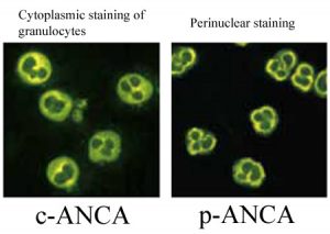
ANCA tests are mostly performed by indirect immunofluorescence microscopy (IFA). Serum samples are mixed with neutrophils so that they can react with autoantibodies. Then, the sample is spread on a glass (slide) and fixed with ethanol, stained with fluorescently labeled anti-human immunoglobulins and examined under a fluorescent microscope and the image patterns are recorded. There are three image patterns mainly c-ANCA, p-ANCA and a-ANCA:
- The cytoplasmic pattern is associated with PR3 antibodies and is called cANCA, corresponding to diffuse coarse granular cytoplasmic staining.
- The perinuclear pattern is associated with MPO antibodies and is called pANCA. Staining occurs around the nucleus.
- Sometimes there is also the presence of a third pattern, called atypical ANCA (a-ANCA), which does not match both patterns. It shows fine mottled, linear or other cytoplasmic staining unlike other patterns.
Atypical ANCA is an example where a clear distinction between cytoplasmic and peripheral staining cannot be made in indirect immunofluorescence technique. It develops against antigens such as cathepsin G, elastase, and lactoferrin. It can be seen in rheumatoid arthritis, drug-induced vasculitis and inflammatory bowel disease, chronic infections.
Fixation of neutrophils with cross-linking fixators such as formalin is recommended, as the simultaneous presence of ANA may exhibit a similar appearance to p- or α-ANCA in ethanol-fixed neutrophils. ANA shows nuclear staining in formalin-fixed neutrophils, whereas p- or α-ANCA shows predominantly cytoplasmic staining.
ELISA method is important in determining ANCA specific antigens (such as PR3, MPO and catalase, lysozyme, elastase, cathepsin). Performing ANCA determination in patients with both indirect immunofluorescence and ELISA and evaluating the result together will increase sensitivity and specificity.
When is ANCA test requested?
These tests are requested in the presence of symptoms and findings suggestive of systemic autoimmune vasculitis in patients. There are non-specific symptoms in the early stages of the disease;
As the disease progresses, symptoms including various complications arise due to damage to the vessels;
These tests can also be used for periodic follow-up in patients diagnosed with systemic autoimmune vasculitis. They can also be used in the differentiation of inflammatory bowel diseases (IBD).
Assessment of ANCA Tests
ANCA test results should be evaluated very carefully, taking into account other laboratory test results, imaging results, and patient symptoms.
If the ANCA test is positive, the next step is to determine the amount of antibody present in the blood, called antibody titer. For this, serum samples are diluted at certain rates and the presence of antibodies is measured at each dilution. The highest dilution at which the presence of antibody is detected is given as the antibody titer result. Results below 1/20 titer are considered negative (Negative: less than 1/20 titer)