


Desmoglein 1 Test / Desmoglein 3 Test
Desmoglein 1 Test / Desmoglein 3 Test, which is the determinant of pemphigus vulgaris, is being studied in our laboratory.
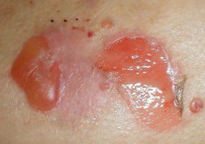
Pemphigus is an immune system disease characterized by the formation of bullae on the skin and mucous membranes.
Pemphigus is one of the diseases that occurs after the immune system fights against one’s own tissues and cells.
Pemphigus, which is not contagious, is an important skin disease that occurs on the skin and mouth, but sometimes in the nose, throat, eyes and genitals, which starts as burn-like blisters filled with liquid and progresses as superficial wounds that burst and open in a short time.
The most important finding of pemphigus vulgaris is the detection of IgG type autoantibodies against the surface of keratinocyte cells. All patients with pemphigus vulgaris have anti desmoglein 3 ( AD3 ) antibodies and some of these patients also have anti desmoglein 1 ( AD1 ) antibodies. Desmoglein 1 and 3 Test, which is the determinant of pemphigus vulgaris, is being studied in our laboratory.
Why Desmoglein 1 Test and Desmoglein 3 Test Are Requested
The screening test of choice (pemphigus) for patients suspected of having an autoimmune blistering disorder of the skin or mucous membranes is used as an aid in the diagnosis of pemphigus.
Method Name
Enzyme-Linked ImmunoSorbent Assay (ELISA)
Where to Work in Istanbul
Istanbul Laboratories Kadıköy and Şişli Branches Tel : 4447522
Reporting Name
Desmoglein 1 and 3, Serum
Method Description
This enzyme-linked immunosorbent assay (ELISA) method detects and measures serum levels of some pemphigus diseases. Calibrators and patient sera are added to microwells coated with DSG1 and DSG3 antigens, allowing the antibodies to react with the immobilized antigens. After washing to remove unbound serum proteins, horseradish peroxidase conjugated IgG is added and incubated. After another wash step, the peroxidase substrate is added and allowed to incubate for an additional time. Stop solution is added to each well to cancel the enzyme reaction and stabilize the color development. The assay can be measured photometrically by measuring the reaction and plotting the results. (Ishii K, Amagai M, Hall RP, et al: Characterization of autoantibodies in pemphigus using antigen-specific ELISA) Co-expresses recombinant desmogleins with Baculovirus. J Immunol 1997 Aug 15; 159 [4]: 2010-2017)
PDF Report
You can get it by Email or Online
Day(s) and Time(s) done
Study once a week on Mondays. (Blood should be given in the laboratory by Friday.)
Runtime
2 days (Samples tested 2 days / week) Result Wednesday 18:00. Maximum Laboratory Time 10 days. Sample Retention Period 14 days.
Test Usage
As an aid in the diagnosis of pemphigus, the screening test of choice (pemphigus) for patients suspected of having an autoimmune blistering disorder of the skin or mucous membranes.
Clinical Information
Pemphigus includes a group of autoimmune, blistering diseases characterized by intraepithelial lesions, often fatal. Pemphigus vulgaris and its variants may occur alone or with mucosal and skin lesions and oral or mucosal lesions. Pemphigus foliase and its variants are present with skin lesions alone.
Indirect immunofluorescence (IIF) studies reveal that both forms of pemphigus result from autoantibodies against cell surface antigens of the stratified epithelium or mucous membranes and skin. These antibodies bind to calcium-dependent adhesion molecules at cell surface desmosomes, particularly desmoglein 1 (DSG1) in pemphigus foliaceus and desmoglein 3 (DSG3) and/or DSG1 in pemphigus vulgaris. Desmogleins are protein substances found on and on the surface of keratinocytes. These proteins have been shown to be a critical factor in cell-cell adhesion. Antibodies to Desmogleins can cause loss of cell adhesion, which is the primary cause of blister formation in pemphigus.
The diagnosis of pemphigus depends on biopsy and serum studies that characterize the lesions and detect the autoantibodies that cause them. Initially, serum studies were performed by IIF using monkey esophagus and other tissue substrates. The identification of reactive antigens as DSG1 and DSG3 has made it possible to develop highly specific and sensitive enzyme-linked immunosorbent assay methods.
Comment
Desmoglein 1 (DSG1) and desmoglein 3 (DSG3) antibodies have been shown to be present in patients with pemphigus. Many patients with pemphigus foliaceus, a superficial form of pemphigus, have antibodies to DSG1. Patients with pemphigus vulgaris, a deeper pemphigus, have antibodies to DSG3 and sometimes to DSG1.
Antibody titer correlates in a semiquantitative relationship with disease activity in many patients. Patients with severe disease can generally be expected to have high titers of antibodies to DSG. Titrations are expected to decrease with clinical improvement.
Our experience shows that there is a very good correlation between DSG1 and DSG3 results and the presence of pemphigus. Adequate sensitivity and specificity for disease have been documented. However, it is recommended to use the IIF test (CIFS / Cutaneous Immunofluorescent Antibodies [IgG], Serum) in patients who have pemphigus with clinical findings or routine biopsy and whose DSG test is negative.
Attention
Desmoglein 1 (DSG1) and desmoglein 3 (DSG3) results are diagnostic aids only and should not be interpreted as diagnostic by themselves. Results should be interpreted in conjunction with the patient’s clinical assessment, along with other diagnostic procedures.
The performance of these tests in the pediatric population has not been established. Assay performance characteristics for matrices other than serum have not been established. A positive result indicates the presence of antibodies to recombinant DSG1 and DSG3 and does not specifically identify a specific type of pemphigus. A negative result does not exclude the presence of pemphigus.